Using digitized photographs of chromosomes from real human genetic studies, this exercise simulates the process of karyotyping a human being. Just as if you were working in a genetic analysis program at a hospital or clinic, you will be assembling chromosomes into a finished karyotype and analyzing your results. Over 400,000 kyotype analyses are conducted annually in the United States and Canada. Consider conducting these assessments for actual people, knowing that the results will have a significant impact on their life.
The 23 pairs of human chromosomes condense during mitosis and can be seen under a light microscope. In order to perform a karyotype analysis, cells in mitosis are typically blocked and the condensed chromosomes are stained with Giemsa dye. Chromosome areas rich in the base pairs Adenine (A) and Thymine (T) are stained by the dye, resulting in a black band. It’s a frequent misperception that bands represent individual genes, yet even the smallest bands can include hundreds of genes and over a million base pairs. For instance, the size of one tiny band is about equivalent to the entirety of one bacterium’s genetic code.
The comparison of chromosomes’ lengths, centromere positions (regions where the two chromatids are connected), and G-band positions and sizes is part of the analysis. Three karyotypes will be electronically completed, and you’ll search for anomalies that might explain the phenotypic.
Your homework
Contents
- 1 Definition
- 2 Karyotype
- 3 Chromosomal abnormalities and kyotyping
- 4 Karyotype Creation from Mitotic Cells
- 5 Image of Biology Karyotype Worksheet Answers Key
- 6 Free Download Biology Karyotype Worksheet Answers Key
- 7 Banding Patterns Show Chromosome Structure Specifications
- 8 Some pictures about 'Biology Karyotype Worksheet Answers Key'
- 9 Related posts of "Biology Karyotype Worksheet Answers Key"
The goal of this exercise is to provide a basic understanding of human genetic research. Karyotyping is one of several methods that we can use to search for the thousands of potential hereditary illnesses in people.
You will examine the karyotypes of three patients, complete their case histories, and identify any extra or missing chromosomes. The next step is to search the internet for websites that address various aspects of human genetics. If this is a class assignment, you must submit a total of 7 written responses (2 for each patient, 1 for the internet search).
Definition
An individual’s entire set of chromosomes is known as their karyotype. The phrase can also refer to an image created in a lab showing a person’s chromosomes separated from one cell and organized in numerical order. A karyotype can be used to check for chromosomal number or structural problems.
Karyotype
One of the characteristics of each species is its karyotype. The chromosomes from one cell are photographed, cut out, and arranged into a karyotype utilizing the centromere positions, banding pattern, and size as markers. Under a light microscope, an organism’s morphology and chromosome count are described as its karyotype. Cytogenetic studies include the generation of karyotypes and their investigation. The autosomal chromosomes are present in two copies in normally diploid organisms. Karyotypes can be employed for a variety of things, such as malignancy research or chromosomal iteration studies in prenatal diagnosis. Additionally, to comprehend cellular function, taxonomic relationships, and to provide details on previous evolutionary occurrences. One pair of sex chromosomes and 22 pairs of autosomal chromosomes make up the average human karyotype.
Chromosomal abnormalities and kyotyping
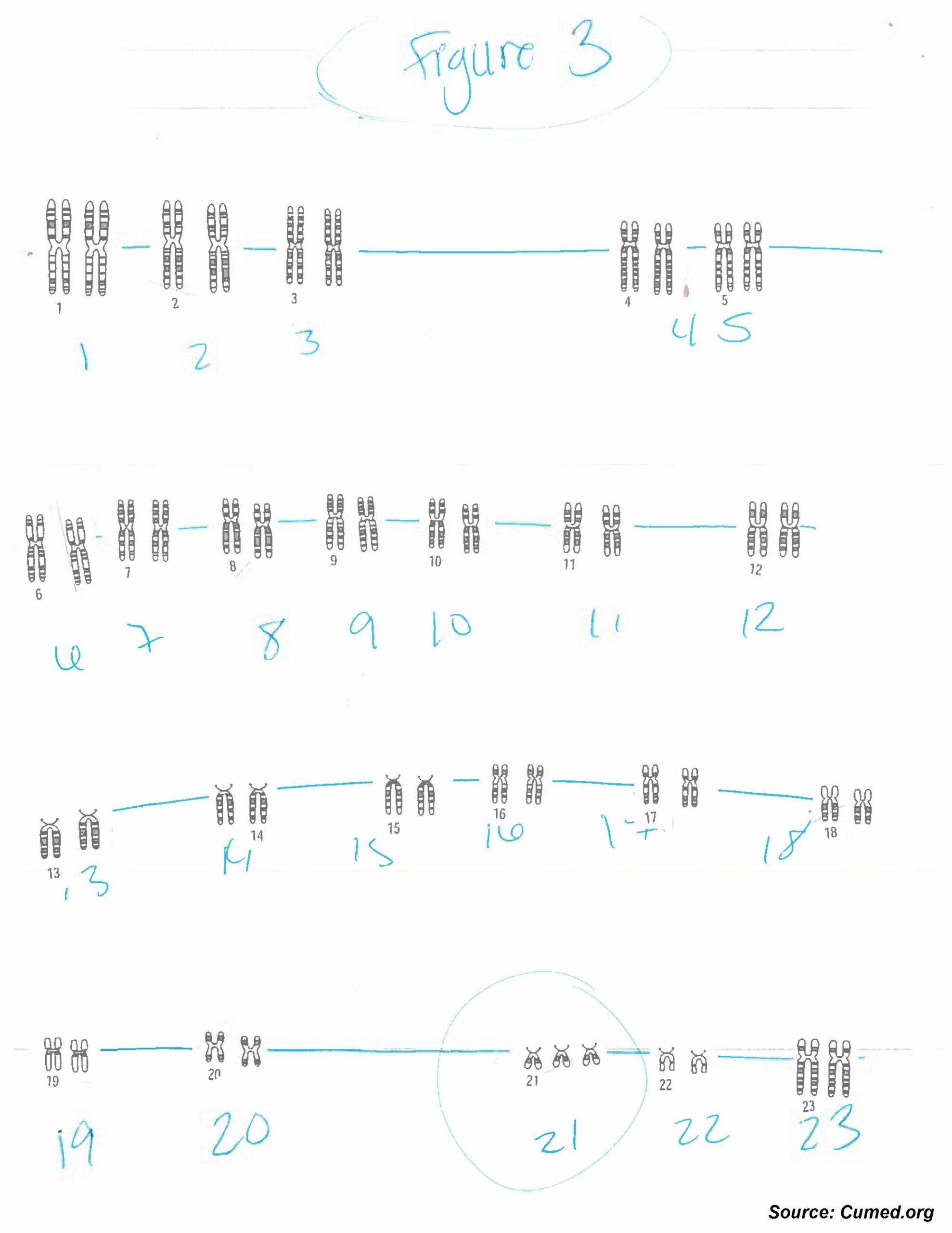
Karyotyping, which pairs and arranges every chromosome in an organism, produces a genome-wide snapshot of a specific person’s chromosomes. Standardized staining techniques are used to create kyotypes, which reveal distinctive structural characteristics for each chromosome. Clinical cytogeneticists examine karyotypes of humans to look for large-scale genetic anomalies—disorders affecting many megabases or more of DNA. Karyotypes can show chromosome number variations linked to aneuploid disorders, like trisomy 21. (Down syndrome). Chromosomal deletions, duplications, translocations, or inversions are examples of more subtle structural abnormalities that can be identified through careful examination of karyotypes. In reality, as medical genetics and clinical medicine grow more closely entwined, karyotypes are turning into a source of diagnostic data for particular birth abnormalities, hereditary diseases, and even malignancies.
Karyotype Creation from Mitotic Cells
Karyotypes are created from mitotic cells that have experienced a cell cycle arrest during the metaphase or prometaphase phase, when chromosomes are in their most compact configurations. These cells can be obtained from a range of tissue types. Tumor biopsies or bone marrow samples are common specimens used to diagnose malignancy. Karyotypes are frequently produced from skin biopsies or peripheral blood samples for various diagnoses. Amniotic fluid or chorionic villus specimens are employed as the cell sources for prenatal diagnostics.
The short-term cultivation of cells obtained from a specimen is the first step in the process of creating a karyotype. Following a period of cell growth and multiplication, the injection of colchicine, which poisons the mitotic spindle, causes dividing cells to be halted in metaphase. Next, a hypotonic solution is applied to the cells, causing their nuclei to enlarge and the cells to explode. The nuclei are then treated with a chemical fixative, placed on a glass slide, and stained in order to reveal the chromosomes’ structural characteristics.
Image of Biology Karyotype Worksheet Answers Key
Free Download Biology Karyotype Worksheet Answers Key
Free Download Biology Karyotype Worksheet Answers Key: Click Here
Banding Patterns Show Chromosome Structure Specifications
Chromosome structural details are difficult to see under a light microscope in their natural state. Therefore, cytologists have created stains that interact with DNA and produce distinctive banding patterns for different chromosomes in order to make analysis more effective and efficient. Chromosome separation was extremely challenging before the invention of these bands methods, therefore they were simply grouped based on size and where their centromeres were located.
When Torbjorn Caspersson and his coworkers introduced the first banding method, known as Q-banding, in 1970, this situation altered. The fluorescent dye quinacrine, which is used in Q-banding and is used to alkylate DNA, is sensitive to quenching over time. Quinacrine produced distinctive and repeatable banding patterns for individual chromosomes, as shown by Caspersson et al. Since then, other alternative chromosomal banding methods have been created, mainly replacing Q-banding in clinical cytogenetics. Giemsa dye is now used to stain the majority of karyotypes, which allows for improved band resolution, a more stable preparation, and analysis using standard bright-field microscopy.
The base composition of the DNA and regional variations in chromatin structure are two of the many molecular factors that contribute to staining variances along a chromosome’s length. The Giemsa staining method known as G-banding, which is most frequently employed in North America, involves temporarily treating metaphase chromosomes with the protein-degrading enzyme trypsin before Giemsa staining. Some chromosomal proteins are partially digested by trypsin, which loosens the chromatin structure and makes the DNA accessible to the Giemsa dye.
In general, heterochromatic areas stain more intensely in G-banding because they typically include AT-rich DNA and few genes. Contrarily, less compacted chromatin integrates less Giemsa stain, and these regions show up as light bands in G-banding. This chromatin is typically GC-rich and more transcriptionally active. Most significantly, G-banding creates patterns that are replicable for each chromosome and that are shared by all members of a species. Figure 1a displays an illustration of human chromosomes stained with Giemsa as they might appear under a microscope. Giemsa staining often results in 400–800 bands spread throughout the 23 pairs of human chromosomes. A G-band is equivalent to between several million and 10 million base pairs of DNA, which is a length of DNA long enough to hold hundreds of genes.